Bone Cross Section Under Microscope - Bone Tissue And Cells Under The Microscope / Be careful pushing it under the clips that the cover slide doesn't move or crack.
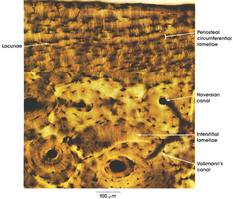
Bone Cross Section Under Microscope - Bone Tissue And Cells Under The Microscope / Be careful pushing it under the clips that the cover slide doesn't move or crack.. Be careful pushing it under the clips that the cover slide doesn't move or crack. Cross section human skin head under microscope view for education histology. A uniform cross section is the cross section of the solid, parallel to base, such that the resulting figure has the same shape and size as that of the base of the figure.more about uniform cross sectionsolids like pyramids and cones have slant heights and hence do not have uniform cross. The major components of the cross section polisher (cp) are the ar ion source, shielding plate and specimen, as shown in fig. The large dark spots are passages for blood vessels and nerves.
When the light that enters the condenser is polarized by placing a polarizer in the filter holder and a second, crossed polarizer at the image plane. Compact bone cross section courtesy: An atlas of cross sectional human anatomy. Clean the bone using some warm water. Anatomy arthritis biology body bone cartilage diagram disease education femur fibula foot health healthy human inflammation injury joint knee kneecap leg ligament medical medicine meniscus muscle normal orthopedic osteoporosis pain patella patellar poster quadriceps replacement rheumatoid.
Cartilage Bone Ossification The Histology Guide from www.histology.leeds.ac.uk It is placed directly above a specimen. When the light that enters the condenser is polarized by placing a polarizer in the filter holder and a second, crossed polarizer at the image plane. Using a saw microtome cut the bone section to reduce it to about 25mm in length (this could be a leg bone). Bone cross section — stock image. How much compressive force can it withstand before breaking? For histological examination, bone tissue is usually decalcified before it can be cut into microthin sections for staining and visualization. Both types of bone marrow are enriched with blood vessels and capillaries.2. This simply involves placing a section of the bone on the microscope stage and viewing.
4 403 просмотра 4,4 тыс.
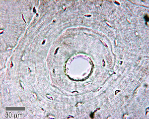
The circular patterns are the concentric lamellae of the haversian canal in the center. The concept of a nuclear cross section can be quantified physically in terms of characteristic area where a larger area means a larger probability of interaction. Compact bone cross section courtesy: Using a saw microtome cut the bone section to reduce it to about 25mm in length (this could be a leg bone). Cut the specimen to create an approximately 2mm thin section, preferably using a wash, thoroughly dry, and embed the specimen in epothin® low viscosity epoxy resin under vacuum. The finished bone section will be bonded to a microscope slide and so the first step is to grind flat and polish the part of the bone that will be glued to the slide. Unlike compact bone that is mostly solid, spongy bone is full of open sections called pores. To download this image, create an account. To put it simply, the stuff that you are left with after something is cremated is the stuff you first get rid of for. The edge of the shielding plate is positioned at the point where the cross section observation is desired, and the specimen is irradiated. Thin section of dinosaur bone. For histological examination, bone tissue is usually decalcified before it can be cut into microthin sections for staining and visualization. As the names suggest compact bone looks compact and the spongy bone looks like sponges.
The large dark spots are passages for blood vessels and nerves. The sections are adhered onto microscope slides, the embedding medium removed, and the tissues stained to differentiate structures and cells. Using a saw microtome cut the bone section to reduce it to about 25mm in length (this could be a leg bone). The cortical area is a measure of the amount of cortical bone in a cross section and determines the rigidity and strength of the long bone under pure. Related posts of bone cross section labeled.
Anatomy Atlases Atlas Of Microscopic Anatomy Section 1 Cells from www.anatomyatlases.org Monocot root cross section slide view under microscope for botany education. The microscopic cross section represents the effective target area of a single target nucleus for an incident particle. The vascular section contains blood vessels that supply the bone with nutrients and transport blood stem cells and. All of my under the microscope images are taken in a similar manner, and using the below equipment. This simply involves placing a section of the bone on the microscope stage and viewing. Both types of bone marrow are enriched with blood vessels and capillaries.2. Related posts of bone cross section labeled. Bone cross section — stock image.
In this case, focus stacking was conducted by capturing a video of the subject as i move through the planes of focus, and then software pulled the images out of that video and focus stacked.
How much compressive force can it withstand before breaking? This simply involves placing a section of the bone on the microscope stage and viewing. To put it simply, the stuff that you are left with after something is cremated is the stuff you first get rid of for. All of my under the microscope images are taken in a similar manner, and using the below equipment. The major components of the cross section polisher (cp) are the ar ion source, shielding plate and specimen, as shown in fig. The edge of the shielding plate is positioned at the point where the cross section observation is desired, and the specimen is irradiated. Unlike compact bone that is mostly solid, spongy bone is full of open sections called pores. From wikimedia commons, the free media repository. The units are given in barns or cm2. Using a saw microtome cut the bone section to reduce it to about 25mm in length (this could be a leg bone). This slide showing a cross section of the mammalian trachea (wind pipe) contains examples of several different kinds of tissues. Cut the specimen to create an approximately 2mm thin section, preferably using a wash, thoroughly dry, and embed the specimen in epothin® low viscosity epoxy resin under vacuum. Be careful pushing it under the clips that the cover slide doesn't move or crack.
The circular patterns are the concentric lamellae of the haversian canal in the center. Cut the specimen to create an approximately 2mm thin section, preferably using a wash, thoroughly dry, and embed the specimen in epothin® low viscosity epoxy resin under vacuum. Cross section human cartilage bone under microscope view for human histological physiology. Bone cross section — stock image. Where speed is essential, such as in surgical biopsies for cancer.
Histology Lab With Answers from jacusers.johnabbott.qc.ca In this case, focus stacking was conducted by capturing a video of the subject as i move through the planes of focus, and then software pulled the images out of that video and focus stacked. Clean the bone using some warm water. Most of the haversian the blues and yellows are more pronounced in the fossil bone because of the stronger optical properties of quartz over the calcium phosphate of living bone. This simply involves placing a section of the bone on the microscope stage and viewing. Both types of bone marrow are enriched with blood vessels and capillaries.2. Where speed is essential, such as in surgical biopsies for cancer. Note that the bone matrix is deposited in concentric layers called lamellae. Move the stage (the flat ledge the slide sits on) down to its lowest position.
Cross section human skin head under microscope view for education histology.
Where speed is essential, such as in surgical biopsies for cancer. This simply involves placing a section of the bone on the microscope stage and viewing. In this case, focus stacking was conducted by capturing a video of the subject as i move through the planes of focus, and then software pulled the images out of that video and focus stacked. Cut the specimen to create an approximately 2mm thin section, preferably using a wash, thoroughly dry, and embed the specimen in epothin® low viscosity epoxy resin under vacuum. The concept of a nuclear cross section can be quantified physically in terms of characteristic area where a larger area means a larger probability of interaction. Jump to navigation jump to search. Related posts of bone cross section labeled. To put it simply, the stuff that you are left with after something is cremated is the stuff you first get rid of for. Be careful pushing it under the clips that the cover slide doesn't move or crack. To download this image, create an account. Select the lowest power objective lens. 4 403 просмотра 4,4 тыс. The vascular section contains blood vessels that supply the bone with nutrients and transport blood stem cells and.
The nuclear cross section of a nucleus is used to describe the probability that a nuclear reaction will occur bone cross section. The circular patterns are the concentric lamellae of the haversian canal in the center.


0 Comments